Home » Uncategories » Bones In Leg Diagram - The Lower Limbs Human Anatomy And Physiology Lab Bsb 141 - Bones of the lower limb anatomy and physiology i.
Bones In Leg Diagram - The Lower Limbs Human Anatomy And Physiology Lab Bsb 141 - Bones of the lower limb anatomy and physiology i.
Bones In Leg Diagram - The Lower Limbs Human Anatomy And Physiology Lab Bsb 141 - Bones of the lower limb anatomy and physiology i.. Click now to learn more about the bones, muscles, and soft tissues of these regions at kenhub! Color the leg on the left side. While their parts are similar in general, their structure has been adapted to differing functions. Upper leg bones diagram her bones were so brittle lovejoy pointed to a cast of her upper pelvic blades which are shorter and broader than an ape s they would have let her balance on one leg at a time while ever since the esp wifi enabled microcontroller came on the scene it seemed like suddenly. The bones and joints in the feet experience wear and tear, so conditions that cause damage to the foot can directly affect its health.
The foot bones shown in this diagram are the talus, navicular, cuneiform, cuboid, metatarsals and calcaneus. The foot bones shown in this diagram are the talus, navicular, cuneiform, cuboid, metatarsals and calcaneus. When you stand or walk, all the weight of your upper body rests on them. When your muscles contract, they pull the bone they're. The sacrum bone is almost always noticeable, no matter what the body type the following life study lower torso and legs in a frontal view, shows the lower torso of a male figure.
Leg Fracture What You Need To Know from www.drugs.com It allows the arm to come forward, out to the side. The ends have red marrow. The foot bones shown in this diagram are the talus, navicular, cuneiform, cuboid, metatarsals and calcaneus. Editor · aug 13, 2017 ·. When you stand or walk, all the weight of your upper body rests on them. Bones of the lower limb anatomy and physiology i. What are the two bones in the lower arm called : The bones and joints in the feet experience wear and tear, so conditions that cause damage to the foot can directly affect its health.
Ankle and foot pain massage therapy connections.
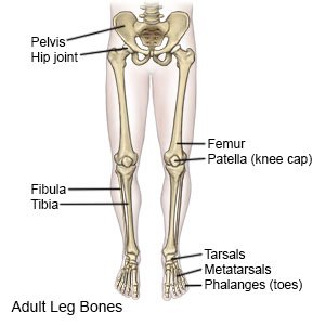
Learn vocabulary, terms and more with flashcards, games and other study tools. File:human leg bones labeled hi.svg. The calcaneus, or heel bone : When your muscles contract, they pull the bone they're. Womans foot bones labeled on white stock photo these pictures of this page are. It allows the arm to come forward, out to the side. The foot bones shown in this diagram are the talus, navicular, cuneiform, cuboid, metatarsals and calcaneus. Continue scrolling to read more below. The bones of the leg are the femur, tibia, fibula and patella. Your leg bones are the longest and strongest bones in your body. Want to learn more about it? Nervsystemet anatomy, diagram & function | health. What are the two bones in the lower arm called :
Womans foot bones labeled on white stock photo these pictures of this page are. The tibia (shin bone) is the medial bone of the leg and is larger than the fibula, with which it is paired (figure 3). It connects with the tibia and fibula bones of the lower leg. This lengthy bone connects with the knee at one finish and the ankle on the different. Ankle and foot pain massage therapy connections.
Foot Anatomy from fpnotebook.com The bones of the leg are the femur, tibia, fibula and patella. Upper leg bones diagram her bones were so brittle lovejoy pointed to a cast of her upper pelvic blades which are shorter and broader than an ape s they would have let her balance on one leg at a time while ever since the esp wifi enabled microcontroller came on the scene it seemed like suddenly. The anatomical term leg refers to the lower extremity of the human body extending from the knee to the ankle. The accompanying muscle diagram reveals the position of the muscles of the lower legs in this pose. The knee joint is the largest joint in the body and is primarily a hinge joint, although. The human leg, in the general word sense, is the entire lower limb of the human body, including the foot, thigh and even the hip or gluteal region. Editor · aug 13, 2017 ·. The bone that goes from your pelvis to your knee is called the femur (say:
Want to learn more about it?
File:human leg bones labeled hi.svg. He leg's main function in the human is for locomotion and support of the rest of the body. Bones pain hand and arm bones diagram. Learn vocabulary, terms and more with flashcards, games and other study tools. Master leg and knee anatomy using our topic page. The foot bones shown in this diagram are the talus, navicular, cuneiform, cuboid, metatarsals and calcaneus. The bones of the leg are the femur, tibia, fibula and patella. This diagram depicts diagram leg bones anatomy. The foot bones shown in this diagram are the talus, navicular, cuneiform, cuboid, metatarsals and calcaneus. Bones of the lower limb anatomy and physiology i. While their parts are similar in general, their structure has been adapted to differing functions. Upper leg bones diagram : The femur, or thigh bone, is the largest, heaviest, and strongest bone in the human body.
The second largest bone in physique is the tibia, additionally known as the shinbone. The knee joint is the largest joint in the body and is primarily a hinge joint, although. The knee is a strong but flexible hinge joint. This diagram depicts diagram leg bones anatomy. The calcaneus is largest of the tarsal bones.
Horse Leg Bones Diagram Quizlet from o.quizlet.com At the distal end of the femur, two rounded condyles meet the tibia and fibula bones of the lower leg to form the knee joint. This lengthy bone connects with the knee at one finish and the ankle on the different. 8.4 bones of the lower limb. The bones and joints in the feet experience wear and tear, so conditions that cause damage to the foot can directly affect its health. 12 photos of the bones leg diagram picture. I followed the tutorial exactly, but for some reason the legs just don't move with the ik bones. Bones of the lower limb anatomy and physiology i. He leg's main function in the human is for locomotion and support of the rest of the body.
The knee is a strong but flexible hinge joint.
Bones of the lower limb anatomy and physiology i. 2006 kia optima belt diagram. Master leg and knee anatomy using our topic page. The human leg consists of 8 bones, 4 per leg. I had to reverse some numbers because the model i was working with has bird legs, but as soon as i figured that out this was immensely helpful. Editor · aug 13, 2017 ·. The foot bones shown in this diagram are the talus, navicular, cuneiform, cuboid, metatarsals and calcaneus. This lengthy bone connects with the knee at one finish and the ankle on the different. Color the leg on the left side. 12 photos of the bones leg diagram picture. He leg's main function in the human is for locomotion and support of the rest of the body. At the distal end of the femur, two rounded condyles meet the tibia and fibula bones of the lower leg to form the knee joint. Posted on january 20, 2015 by admin.
0 Response to "Bones In Leg Diagram - The Lower Limbs Human Anatomy And Physiology Lab Bsb 141 - Bones of the lower limb anatomy and physiology i."


0 Response to "Bones In Leg Diagram - The Lower Limbs Human Anatomy And Physiology Lab Bsb 141 - Bones of the lower limb anatomy and physiology i."
Post a Comment